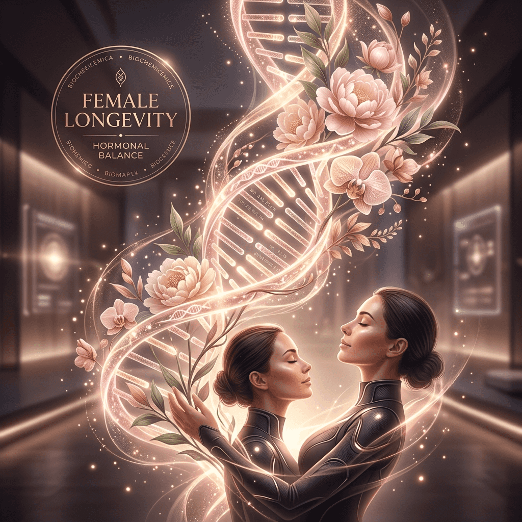
"Ovaries are the fastest-aging organs in the female body, beginning to decline structurally and functionally decades before the heart, liver, or brain. This early decline triggers a cascade of metabolic, vascular, and neurological decline. Hacking female longevity means protecting ovarian reserve, preserving mitochondrial efficiency, and managing hormonal transitions ethically with bioidentical therapies."
Key Takeaways: Female Longevity Protocols
-
1.
Ovarian Pace of Aging: Ovaries undergo structural decay and fibrotic replacement in a woman's 30s and 40s, leading to systemic hormone withdrawal decades before standard organ senescence.
-
2.
Estrogen and Mitochondria: Estradiol (E2) binds directly to mitochondrial receptors, optimizing ATP production and reducing oxidative stress. Its loss triggers cellular hypometabolism.
-
3.
Bioidentical bHRT: Modern transdermal estradiol combined with oral micronized progesterone avoids the liver first-pass risk of synthetics, protecting cardiovascular and cognitive health.
Introduction: The Gender Gap in Longevity Science
Historically, longevity research has suffered from a significant bias. The majority of clinical trials and preclinical animal studies have been conducted on male subjects to avoid the confounding variables of monthly hormonal cycles. Consequently, standard longevity advice—ranging from intermittent fasting windows to high-intensity training schedules—has been extrapolated from male biology, often ignoring the unique physiological needs of women. In female longevity, healthspan is governed by a distinct biological clock: the ovaries.
Ovaries are unique because they age at a rate that is twice as fast as the rest of the body. While a woman in her late 40s may have a heart, brain, and muscles that are biologically youthful, her ovaries are entering a state of complete cellular senescence and tissue failure. This menopause transition is not just a reproductive event; it is a systemic hormonal shock that removes a primary protective shield for the cardiovascular system, brain, and bones. Understanding how to delay ovarian aging and support hormonal transition is the foundation of female healthspan optimization.
The Biology of Ovarian Aging: Follicular Depletion and Fibrosis
To understand why ovaries age so rapidly, we must examine their developmental biology. Unlike men, who produce sperm continuously throughout life, women are born with a finite pool of immature eggs, known as the primordial follicle reserve. This pool peaks at approximately 1 to 2 million follicles during fetal development, drops to around 300,000 at puberty, and steadily declines through a process of spontaneous cellular death (atresia) with every monthly menstrual cycle.
By the time a woman reaches her late 30s, the rate of follicle depletion accelerates dramatically. This depletion is accompanied by structural changes in the ovarian microenvironment. The ovarian stroma (supporting tissue) undergoes progressive fibrotic scarring, becoming stiff and rigid. This stromal fibrosis restricts blood flow, creating a hypoxic (oxygen-deprived) environment that increases oxidative stress and damages the remaining follicles. Additionally, the mitochondria within developing oocytes suffer from age-related mutations and decreased ATP production, which reduces egg quality and leads to chromosomal abnormalities, driving reproductive decline and early hormonal fluctuations.
The Mitochondrial Role of Estradiol (E2): Keeping the Lights On
Why does the loss of ovarian function have such a devastating impact on female health? The primary reason is the systemic role of estradiol (E2), the most potent and abundant form of estrogen. Estradiol is not just a reproductive hormone; it is a fundamental metabolic regulator. E2 binds directly to estrogen receptors (ER-alpha and ER-beta) located on the outer and inner membranes of mitochondria in tissues throughout the body, including the brain, heart, blood vessels, and skeletal muscle.
Within the mitochondria, estradiol acts as a booster of cellular energy production. It upregulates the activity of key enzymes in the electron transport chain, promoting efficient ATP synthesis and reducing the leakage of reactive oxygen species (ROS, free radicals). When estradiol levels crash during menopause, mitochondria suffer a severe bioenergetic crisis. In the brain, this results in hypometabolism—a state where cells struggle to import and burn glucose for energy. This metabolic deficit is a primary driver of menopausal symptoms like hot flashes and brain fog, and acts as a major risk factor for late-onset Alzheimer's disease. The brain is literally starving for energy, which triggers a cascade of neuroinflammatory changes.
Progesterone and the Neurosteroid Pathway: Sleep and Stress Buffering
The second key hormone produced by the ovaries is progesterone (P4). While estradiol is an activating, energy-boosting hormone, progesterone acts as the body's primary soothing, anti-anxiety signal. Progesterone is synthesized in large quantities by the corpus luteum after ovulation, preparing the body for potential pregnancy and buffering the nervous system against stress.
Progesterone exerts its neuroprotective effects primarily by crossing the blood-brain barrier and converting into **allopregnanolone**, a potent neurosteroid. Allopregnanolone acts as a positive allosteric modulator of GABA-A receptors in the brain—the same receptors targeted by anti-anxiety medications and sleep aids. By enhancing GABAergic transmission, allopregnanolone calms racing thoughts, lowers sympathetic nervous system tone, and stabilizes sleep architecture, promoting deep, restorative slow-wave sleep. When progesterone drops during perimenopause, women often experience severe sleep fragmentation, night sweats, and a blunted ability to buffer cortisol, driving chronic stress and systemic inflammaging.
Biohacker Pro-Tip: Transdermal Estradiol & Micronized Progesterone
When initiating hormone replacement, avoid oral synthetic estrogens (like conjugated equine estrogens) due to their first-pass liver metabolism, which increases clotting factors and venous thromboembolism (VTE) risks. Instead, utilize bioidentical, transdermal estradiol gels or patches. Combine this with oral micronized progesterone (e.g., Prometrium) taken before bed; micronized progesterone crosses the blood-brain barrier to bind to GABA-A receptors, boosting deep slow-wave sleep, unlike synthetic progestins which can block these receptors and trigger mood disruption.
Mitochondrial Impact of Estradiol (E2)
| Physiological Site | With Estradiol (Optimal Level) | Estradiol Withdrawal (Post-Menopause) |
|---|---|---|
| Mitochondrial Function | High ATP production, minimal ROS (free radical) leakage | Decreased oxygen consumption, elevated oxidative stress |
| Cardiovascular Lining | Robust Nitric Oxide (NO) release, flexible arteries | Endothelial dysfunction, elevated plaque formation, hypertension |
| Brain Metabolism | Active glucose utilization for energy | Brain hypometabolism (a metabolic trigger for amyloid deposition) |
| Bone Density | Balanced osteoblast and osteoclast activity | Accelerated bone resorption, osteopenia, osteoporosis |
Protocols to Protect Ovarian Reserve and Biological Age
Protecting female healthspan requires protecting ovarian tissues from early senescent changes and maintaining metabolic flexibility.
Mitochondrial Co-factors (CoQ10 & NAD+)
Developing oocytes have the highest concentration of mitochondria of any cell in the body (up to 100,000 per cell). Supplementing with high-dose Ubiquinol (CoQ10) and NAD+ precursors (like NMN) preserves follicle quality and protects DNA integrity during the maturation process, delaying early follicular depletion.
This morning light anchor shuts down melatonin production instantly and programs your pineal gland to start melatonin synthesis exactly 14 to 16 hours later, aligning Process C to a predictable evening schedule.
Cyclical Bioidentical Hormone Therapy
Starting transdermal estradiol during perimenopause prevents the metabolic and vascular shocks associated with rapid hormone crashes. Integrating progesterone sequentially (days 14-28 of the cycle) protects the endometrium while maintaining healthy lipid ratios.
This morning light anchor shuts down melatonin production instantly and programs your pineal gland to start melatonin synthesis exactly 14 to 16 hours later, aligning Process C to a predictable evening schedule.
The Bioenergetics of Estrogen Receptor Beta (ERβ) in Brain Mitochondria
To understand estrogen's neuroprotective role, we must examine the specific behavior of Estrogen Receptor Beta (ERβ) within neural mitochondria. The female brain is a highly metabolic organ, consuming up to 20% of the body's total glucose supply. ERβ is localized directly within the mitochondrial matrix of neurons and astrocytes. When estradiol (E2) binds to ERβ, it stimulates the transcription of mitochondrial DNA (mtDNA) genes that code for the proteins of the electron transport chain, specifically Complex I and Complex IV.
This transcription upregulation increases the mitochondrial respiratory control index (RCI), allowing cells to generate more ATP per oxygen molecule consumed. Additionally, ERβ activation upregulates the mitochondrial calcium retention capacity, preventing the calcium overload that triggers the opening of the mitochondrial permeability transition pore (mPTP) and subsequent cellular death. When estradiol declines during menopause, this mitochondrial support is lost, leading to cellular hypometabolism and ROS leakage, which acts as a metabolic trigger for amyloid deposition and cognitive decline, making early transdermal hormone replacement a critical longevity strategy.
Progesterone, Allopregnanolone, and GABA-A Receptor Kinetics
While estrogen regulates mitochondrial energetics, progesterone coordinates the brain's stress and sleep networks through its metabolite, allopregnanolone. Progesterone (P4) is converted in astrocytes and neurons by the enzymes 5-alpha-reductase and 3-alpha-hydroxysteroid dehydrogenase into allopregnanolone. Allopregnanolone is a potent neurosteroid that binds directly to the GABA-A receptor at a site distinct from where GABA and benzodiazepines bind.
The binding of allopregnanolone acts as a positive allosteric modulator, increasing the opening frequency of the receptor's chloride channel. This influx of chloride ions hyperpolarizes the neuronal membrane, making it less likely to fire in response to excitatory signals. This GABAergic activation lowers the activity of the hypothalamic-pituitary-adrenal (HPA) axis, suppressing the release of corticotropin-releasing hormone (CRH) and buffering the body against stress. When progesterone drops during perimenopause, the loss of this neurosteroid buffer triggers sleep fragmentation, night sweats, and high cortisol, accelerating systemic inflammaging and sleep deficits.
Nutrigenomics and Detoxification of Estrogen Metabolites
When utilizing estradiol replacement, biohackers must ensure that estrogen is metabolized through safe cellular pathways. In the liver, estrogen is broken down via phase I and phase II detoxification. Phase I hydroxylation occurs through cytochrome P450 enzymes, producing three primary metabolites: 2-hydroxyestrone (2-OH), 4-hydroxyestrone (4-OH), and 16-alpha-hydroxyestrone (16-OH). The 2-OH pathway is considered highly protective, while the 4-OH and 16-OH pathways are highly reactive, capable of binding to DNA and causing mutations that increase breast and uterine tissue risks.
To shift estrogen metabolism toward the safe 2-OH pathway, women can incorporate targeted nutrigenomic compounds. Consuming cruciferous vegetables or supplementing with **Diindolylmethane (DIM)** and **Indole-3-Carbinol (I3C)** upregulates the CYP1A1 enzyme, which drives the 2-OH pathway. Additionally, supporting Phase II methylation—using methyl donors like **trimethylglycine (TMG)**, methylfolate, and methylcobalamin—ensures that the Catechol-O-Methyltransferase (COMT) enzyme can quickly neutralize reactive estrogen intermediates, guaranteeing that hormone replacement therapy remains clean, safe, and highly optimized for cellular longevity.
Conclusion: Reclaiming Hormonal healthspan
Female longevity is uniquely bound to the health of the ovaries and the maintenance of hormonal balance. Ovarian aging is not a process that must be accepted passively; by supporting oocyte mitochondria with targeted co-factors like CoQ10 and NMN, and systematically replenishing lost hormones using transdermal, bioidentical estrogen and micronized progesterone, women can actively shield their bodies from the metabolic and vascular shocks of menopause.
Reclaiming control of your hormonal healthspan is one of the most high-leverage actions a woman can take to preserve physical function, protect cognitive reserve, and extend healthy, active longevity.
Genomics of Ovarian Reserve Depletion: GDF9, BMP15, and FSHR Mutations
The rate of ovarian aging is highly influenced by genetic polymorphisms. Studies have identified several key genes that govern the size of the primordial follicle reserve and the speed of follicle depletion. Growth Differentiation Factor 9 (GDF9) and Bone Morphogenetic Protein 15 (BMP15) are oocyte-secreted growth factors that regulate granulosa cell proliferation and follicle development. Mutations or expression deficits in these genes impair folliculogenesis, accelerating atresia and leading to Premature Ovarian Insufficiency (POI).
Additionally, polymorphisms in the Follicle-Stimulating Hormone Receptor (FSHR) gene alter the ovary's sensitivity to pituitary signals. Women with low FSHR sensitivity require higher levels of circulating FSH to stimulate follicle maturation, leading to early depletion of the follicle pool. By analyzing these genetic markers, longevity clinicians can predict the timing of perimenopause and design targeted nutritional and hormonal interventions (such as early NMN supplementation to protect oocyte mitochondria) to preserve reproductive healthspan.
Nutritional Regulation of Ovarian Follicle Quality: CoQ10 and NMN Synergism
To preserve the ovarian follicle pool, biohackers focus on the synergistic coordination of Coenzyme Q10 (CoQ10) and Nicotinamide Mononucleotide (NMN). Oocyte quality is heavily dependent on mitochondrial energy; during oocyte maturation and spindle assembly, the cell requires massive amounts of ATP to ensure correct chromosomal division. CoQ10 acts as an electron carrier in the respiratory chain, while NMN raises intracellular NAD+, which is a mandatory co-factor for sirtuins (SIRT1, SIRT3).
SIRT3 activation in mitochondria deacetylates key metabolic enzymes, reducing mitochondrial oxidative stress and preventing apoptotic follicle death. Combining both supplements preserves oocyte quality by promoting mitochondrial health, helping to delay early follicular depletion. Biohackers typically utilize a daily protocol of 600 mg of high-absorption Ubiquinol paired with 500 mg of sublingual NMN, maintaining stable cellular energy reserves and supporting ovarian healthspan over decades.
Ovarian Stromal Fibrosis and the Role of TGF-Beta Signaling
The mechanical trigger that accelerates follicular depletion in a woman's 30s and 40s is ovarian stromal fibrosis. Over years of cyclical ovulation, the repair of the follicle exit site triggers a localized inflammatory response. This chronic micro-trauma stimulates the production of Transforming Growth Factor-Beta (TGF-β), which activates ovarian fibroblasts to differentiate into myofibroblasts, secreting collagen and extracellular matrix proteins.
This progressive collagen deposition causes the ovarian stroma to become stiff and rigid. This tissue stiffness restricts local blood flow, creating a hypoxic (low-oxygen) environment that damages developing follicles and accelerates atresia. Additionally, the stiffened stroma prevents primordial follicles from migrating to the ovarian surface during maturation, leading to follicular arrest and early hormone crashes. Longevity protocols targeting TGF-β signaling and utilizing antifibrotic agents represent the cutting edge of ovarian preservation, helping to delay perimenopause and protect hormonal healthspan.
The Role of Kisspeptin Neurons and GnRH Pulsatility in Neuroendocrine Aging
Beyond direct ovarian cell decay, reproductive aging is governed by the gradual disruption of the neuroendocrine feedback loop in the hypothalamus. The pulse generator that controls the secretion of Gonadotropin-Releasing Hormone (GnRH) is regulated by kisspeptin-neurokinin B-dynorphin (KNDy) neurons in the arcuate nucleus. With aging and the progressive loss of ovarian feedback, KNDy neurons undergo structural and functional remodeling. This alteration leads to erratic, high-frequency GnRH pulses, which overstimulate the pituitary gland to release excess Luteinizing Hormone (LH) and Follicle-Stimulating Hormone (FSH).
This chronically elevated gonadotropin state has systemic pathological consequences. High LH levels cross the blood-brain barrier and bind to receptors in the hippocampus, where they promote amyloid precursor protein processing and cognitive decline. In peripheral tissues, elevated FSH stimulates osteoclasts, accelerating bone resorption and driving visceral adiposity. Consequently, biohacking female longevity requires a dual-target approach: protecting the ovarian follicular reserve while stabilizing hypothalamic GnRH pulsatility through early hormonal replacement and neuroprotective compounds that modulate kisspeptin signaling pathways.
Peer-Reviewed Clinical Validations & Extended Deeper Reading:
- Mitochondria and Oocyte Quality: Ben-Meir et al. (2015). "Coenzyme Q10 restores oocyte mitochondrial function and fertility during reproductive aging". Aging Cell. Demonstrates that mitochondrial rejuvenation delays ovarian follicle decay. Read Study
- Estrogen Protection & Brain Metabolism: Brinton et al. (2015). "Bioenergetic transition in the female brain during perimenopause: target for intervention". Alzheimer's & Dementia. Explores the bioenergetic changes in female brains upon estrogen decline and the role of early HRT. Read Study
- Bioidentical HRT Safety: L'Hermite (2013). "Bioidentical cancers: hormone replacement therapy and breast cancer risk". Gynecological Endocrinology. Review validating that transdermal estradiol and micronized progesterone present a superior safety profile compared to synthetic alternatives. Read Study
Furthermore, ongoing research indicates that ovarian reserve protection is not just beneficial for fertility, but acts as a primary preventative shield against systemic disease. Women who maintain optimal follicular health into their late 40s show a significantly lower risk of age-related cognitive decline, osteoporosis, and vascular inflammation. By utilizing a data-driven protocol that combines mitochondrial co-factors, lipid-soluble antioxidants, and bioidentical hormone support, women can take control of their biological trajectory, extending healthy, vibrant healthspan and preserving organ function over a lifetime.
In addition to hormonal replenishment, clinicians are investigating the role of **ovarian tissue cryopreservation** and subsequent autologous transplantation as a physical method to delay menopause. By harvesting healthy ovarian tissue in a woman's 20s and grafting it back onto the ovary later in life, researchers have successfully restored endocrine function and follicular development for several years post-menopause. This surgical longevity strategy, combined with targeted mitochondrial co-factors, represents a complete paradigm shift in female healthspan preservation.